

-
Where the patient lives.
-
Whether they have recently traveled.
-
Whether they have had malaria before.
-
Since when they have had symptoms.
During the physical examination, splenomegaly (enlarged spleen) and hepatomegaly (enlarged liver) are checked.
However, confirmation cannot be made based only on clinical symptoms because the symptoms of malaria can match those of dengue, typhoid, or other febrile illnesses. Therefore, laboratory confirmation is necessary.
Laboratory Diagnosis
Laboratory tests confirm whether the patient has malaria or not, and if so, which species.
1. Microscopic Examination (Gold Standard):
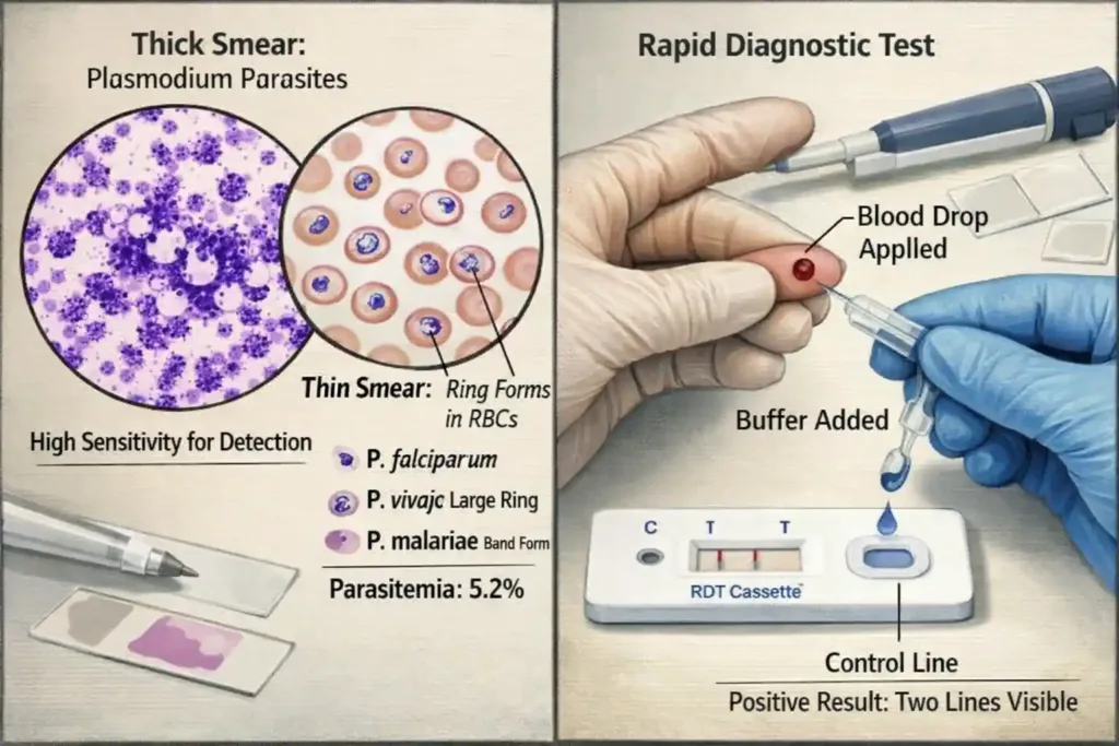
This is the gold standard test for malaria diagnosis. In this, the patient’s blood sample is taken and examined under a microscope.
- Thick Blood Smear:
- The thick smear has a higher concentration of blood, making it easy to detect parasites.
- This is best for screening.
- It tells whether malaria is present or not and what the parasitemia level is (how many parasites are present).
- A drop of blood is placed on a slide, stained with Giemsa stain, and viewed under a microscope.
- Thin Blood Smear:
- The morphology of RBCs is preserved in the thin smear, making species identification easy.
- This confirms which Plasmodium species is present.
- The thin smear shows the exact structure of the parasites, which is different for each species. P. falciparum ring forms, P. vivax enlarged RBCs with Schuffner’s dots, all are identified.
- Parasitemia Count:
- This indicates what percentage of RBCs are infected.
- This assesses the severity of the disease. If parasitemia is more than 5%, it is considered severe malaria, especially in P. falciparum.
- Limitations: Microscopy requires a skilled technician. It is time-consuming (15-30 minutes). If the parasitemia is very low, a false negative result may occur. The quality of staining also affects the result.
2. Rapid Diagnostic Tests (RDTs):
RDTs make malaria diagnosis easy and quick. These kits detect specific antigens of the parasite.
- Principle: RDTs are immunochromatographic tests that detect parasite proteins. The most common antigens detected are:
- HRP-2 (Histidine Rich Protein-2): P. falciparum specific
- pLDH (Plasmodium Lactate Dehydrogenase): Species specific or pan-malarial
- Aldolase: Pan-malarial antigen
- Procedure: It is very simple.
- One or two drops of blood are taken from the patient’s finger prick.
- This is applied to the test kit and Buffer solution is added.
- The result comes in 15-20 minutes.
- The result shows a control line and a test line. The control line confirms that the test is working properly. If the test line is positive, it means the person has malaria.
- Advantages: Very fast (15-20 minutes). No special training or equipment is required. Can be used in remote areas, and is Cost-effective.
- Limitations: Species identification is limited. The parasitemia level is not known. In P. falciparum, HRP-2 can persist for weeks even after the infection is cleared, so a false positive may occur. Very low parasitemia is not detected.
3. Molecular Methods (PCR):
Polymerase Chain Reaction (PCR) is the most sensitive test for malaria.
- Process: This detects the parasite’s DNA. A very small amount of parasite can also be detected. Mixed infections (multiple species simultaneously) are also identified.
- Advantages: Very sensitive (10 times more than microscopy). Can detect low parasitemia. Species identification is accurate. Ideal for research and surveillance.
- Limitations: Expensive. Requires special laboratory equipment. Trained personnel are needed. Time-consuming (several hours). Not practical for routine diagnosis.
PCR is mainly used in research settings, surveillance programs, and complicated cases.
4. Serology (Antibody Detection)
- Use: Checks for malaria antibodies.
-
Purpose: Does not diagnose active infection but indicates past infection. Used for blood donor screening and epidemiological surveys.
Recent Articles

You won’t be fooled again after seeing this

News If a Woman Has Small Breasts, Does It Really Mean Anything About Her Personality or Body? Experts Break Down Common Myths, Social Stereotypes, Biological Facts, and Misconceptions, Revealing Why Body Features Do Not Define Character, Confidence, Health, or Worth, Challenging Outdated Beliefs and Encouraging a More Realistic, Respectful Understanding of Natural Human Diversity and individual differences

Massive python with swollen belly found under family’s shed — X-ray reveals its last meal

THE FIRST ANIMAL YOU NOTICE MAY REVEAL SOMETHING SURPRISING ABOUT YOUR THINKING STYLE

